Labeled Foot Bone Diagram Ct
Foot anatomy bones oblique radiology radiographic ray rays wikiradiography lateral broken xray radiograph schools student look rad right adult ve Ct ankle imaging Common accessory ossicles of the foot
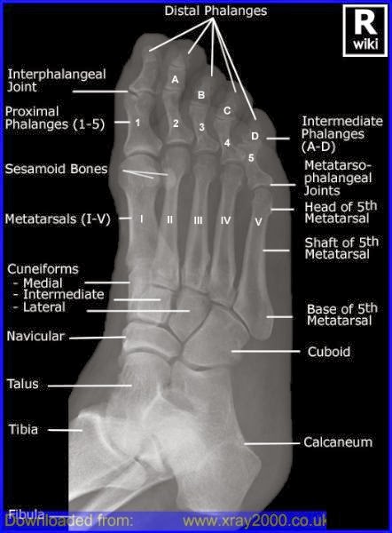
Foot Radiographic Anatomy - wikiRadiography
Foot accessory ossicles radiology common views Foot bones anatomy ankle diagram bone left lower limb skeletal structure lisfranc human feet joint toe physiology anatomie right body Bones of the human foot diagram 1142236 vector art at vecteezy
Radiology oblique bones pedis physiology radiographic annotated imaging wikiradiography xray technology radiologic fracture xrays podiatry nutcracker cuboid coding biomechanics neigong
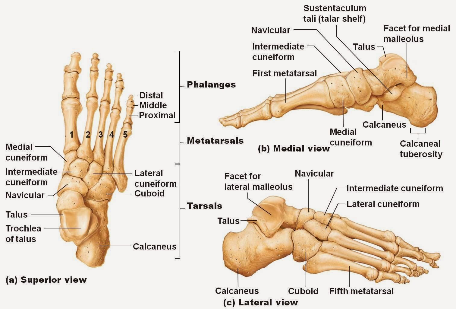
Foot & ankle bonesBones of foot. human anatomy. the diagram shows the placement and names Ankle bones foot labeled calcaneusEmdocs.net – emergency medicine educationcore em: lisfranc injuries.
Bones of the foot diagram imagesOsseous injuries of the foot: an imaging review. part 1: the forefoot Foot anatomy bones diagram human names placement shows alamyBones foot ankle labled labeled separated.

About radiology: teknik radiografi pedis
Forefoot oblique osseous injuries distal emj bmj emermedFoot radiographic anatomy Bones foot anatomy diagram ankle bone human skeletal left feet lower limb physiology body adductus metatarsus joint lisfranc joints labelledFoot bones diagram human vector vecteezy system starting grow.
Common accessory ossicles of the footAccessory ossicles radiology Radiopaedia radiology midfoot fibroma forefootFoot bones x ray / cureus chondromyxoid fibroma of distal phalanx of.

Ankle and foot
.
.


Foot Radiographic Anatomy - wikiRadiography

Common Accessory Ossicles of the Foot | UW Emergency Radiology
About Radiology: Teknik Radiografi Pedis

Bones of the human foot diagram 1142236 Vector Art at Vecteezy

Common Accessory Ossicles of the Foot | UW Emergency Radiology

Calcaneus
Bones of The Foot Diagram images

Bones of foot. Human Anatomy. The diagram shows the placement and names

Foot Bones X Ray / Cureus Chondromyxoid Fibroma Of Distal Phalanx Of